[Most Recent Entries] [Calendar View]
Saturday, August 16th, 2014
| Time | Event | ||||||
| 4:44a | [Herpetology • 2014] Mexican Amber Anole Anolis electrum within A Phylogenetic Context: Implications for the Origins of Caribbean Anoles
Abstract Anoles are well-known examples of adaptive radiation and convergent evolution. Their phylogenetic relationships have been intensely studied, but their fossil record remains fairly poor, limiting our understanding of their evolutionary history. We present new data on Anolis electrum Lazell, 1965, the first discovered fossil anole and sole vertebrate described from Mexican amber, using X-ray computed tomography. We inferred the phylogenetic relationships of A. electrum and comment on its use in estimating the age of Anolis origins, which has significant relevance in explaining the presence of anoles on Caribbean islands. Anolis electrum is represented by two pieces of amber containing parts of the same individual. Partial squamation and skeleton details are well preserved, although only ten characters commonly used in phylogenetic analyses could be scored. The lack of informative characters resulted in A. electrum being inferred in 14 different places within four recognized subclades – Dactyloa, cristatellus series, darlingtoni series, and Norops – one of which corresponds to previously suggested close relationships. Results fail to support a suggested age estimation of 130 Myr for Anolis; consequently, the hypothesis of overwater dispersal as the explanation for the occurrence of anoles on Caribbean islands remains the most robust hypothesis. Keywords: Anolis electrum; Mexican amber; phylogenetics; X-ray computed tomography
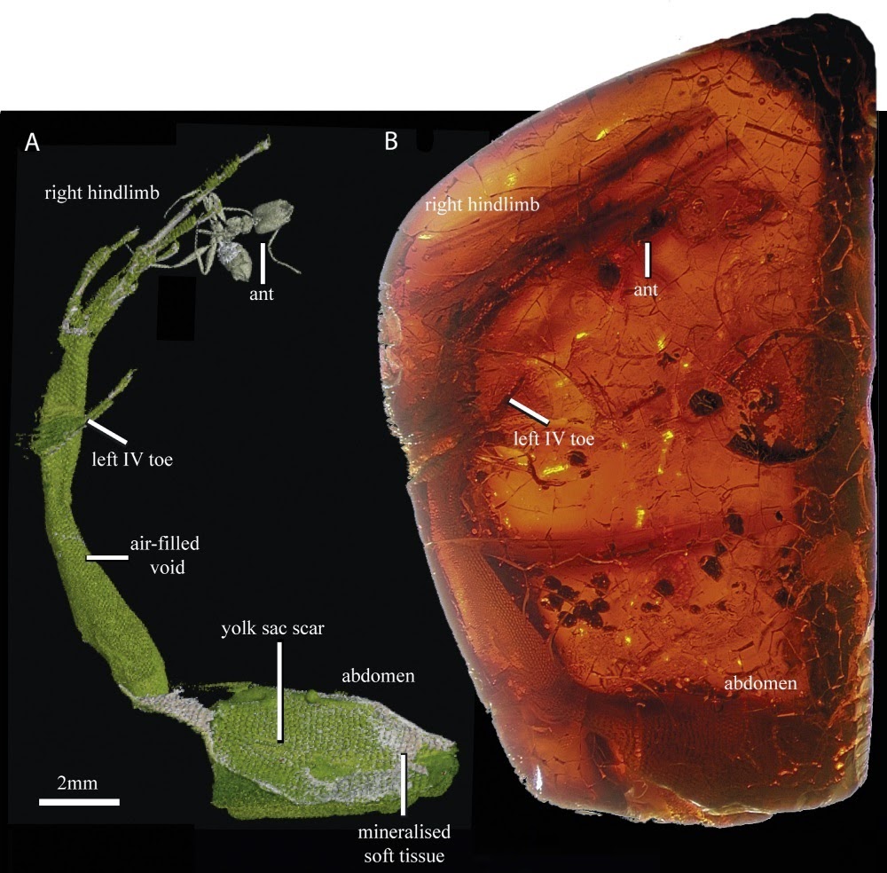
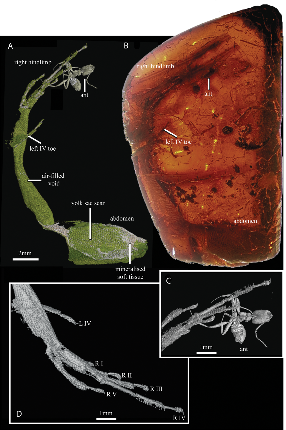
 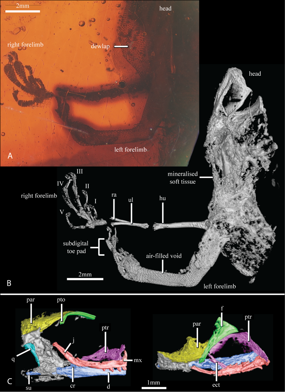 Figure 2. The head, forelimbs, and partial body of Anolis electrum (UCMP 68497), as revealed by light microscopy (A) and high-resolution X-ray computed tomography (HRXCT; B, C). (B) The head and body comprises few skeletal elements obscured by mineralized soft tissue. An air-filled void surrounding the left forelimb reveals scale details from midway along the humerus to the digits. In the right forelimb, the humerus, ulna, radius, metacarpals, and phalanges of all five foretoes are preserved. (C) The skull dissected from the mineralized soft tissue shown in right lateral (left) and dorsal (right) views. For illustration purposes the skull is false-coloured by bone or bone complexes, in which sutures are not visible: green, frontal and postorbital; red, jugal and maxilla; purple, pterygoid and ectopterygoid; blue, dentary, coronoid, and surangular; yellow, parietal; and turquoise, quadrate. Abbreviations: cr, coronoid; d, dentary; ect, ectopterygoid; f, frontal; hu, humerus; j, jugal; mx, maxilla; par, parietal; pto, postorbital bar; ptr, pterygoid; q, quadrate; ra, radius; su, surangular; ul, ulna. | DOI: 10.1111/zoj.12159 María del Rosario Castañeda, Emma Sherratt and Jonathan B. Losos. 2014. The Mexican Amber Anole, Anolis electrum, within A Phylogenetic Context: Implications for the Origins of Caribbean Anoles. Zoological Journal of the Linnean Society. DOI: dx.doi.org/10.1111/zoj.12159 The Fossil Species Anolis electrum Gets an X-ray Makeover anoleannals.org/2014/08/14/the-fossil-sp | ||||||
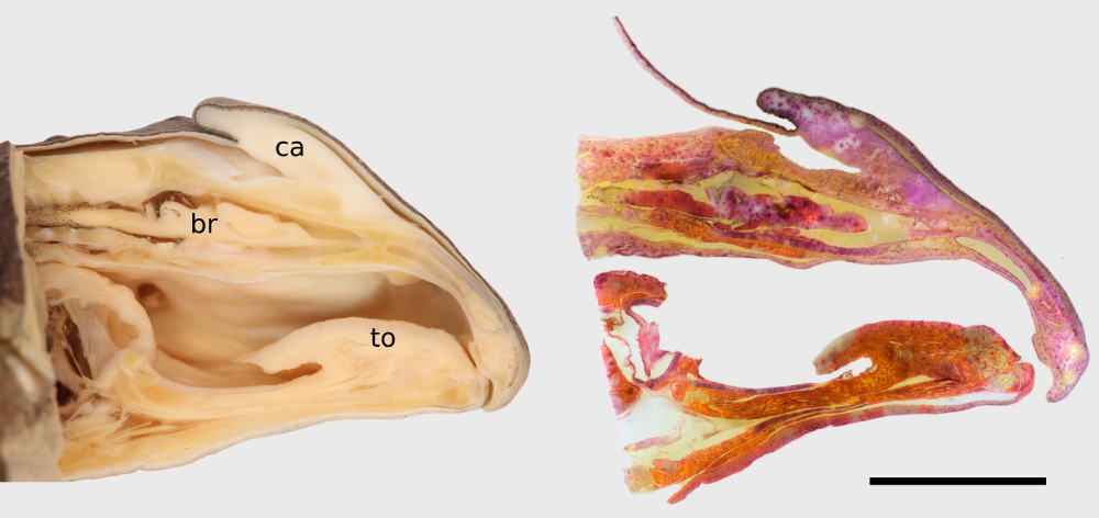
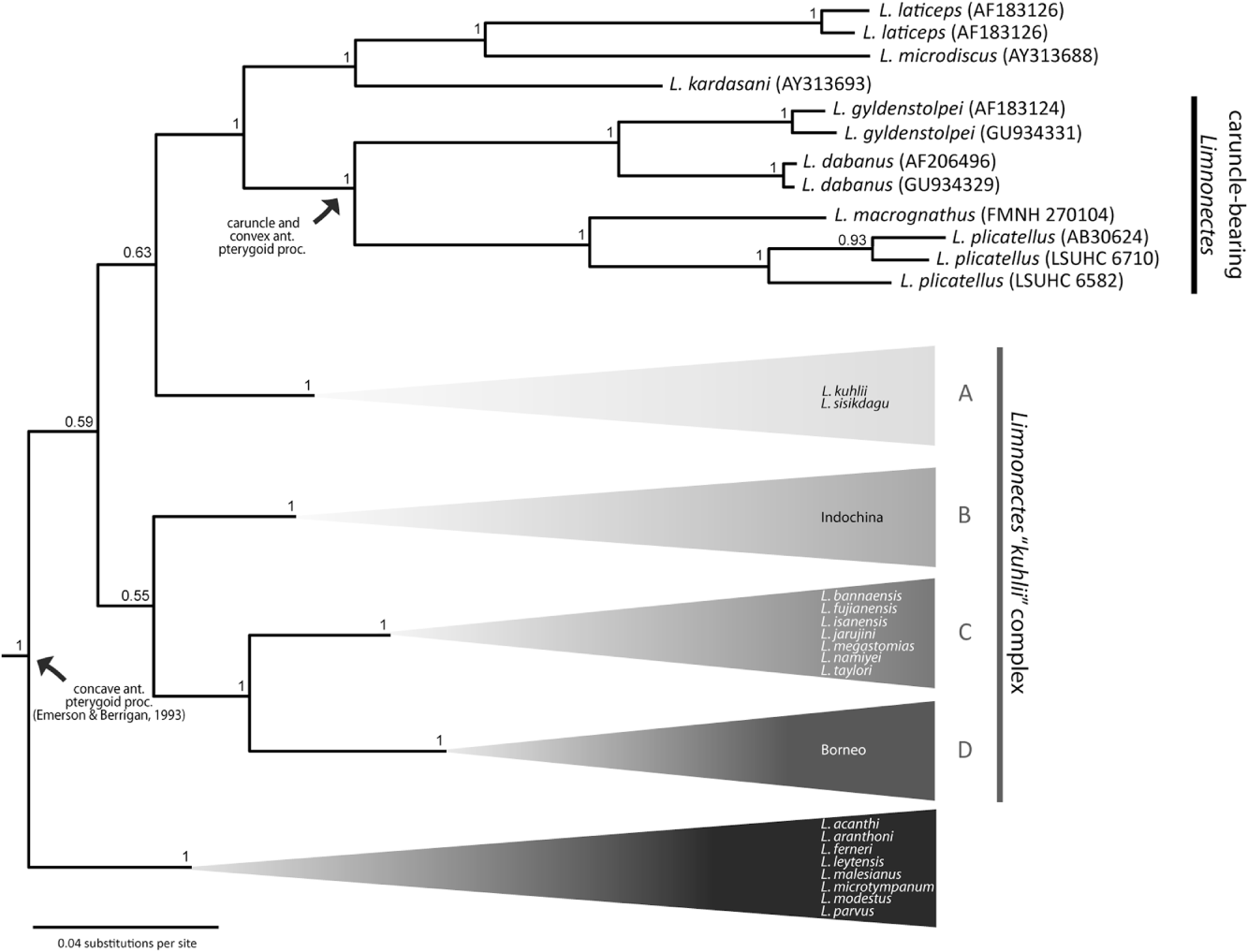
| 6:07a | [Herpetology • 2014] Anatomy, Histology, and Systematic Implications of the Head Ornamentation in the Males of Four Species of Limnonectes (Anura: Dicroglossidae)
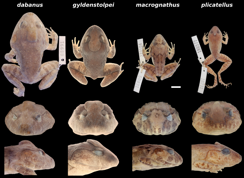
ABSTRACT The males of four species of the Asian frog genus Limnonectes [Limnonectes dabanus (Smith, 1922a), Limnonectes gyldenstolpei (Andersson, 1916), Limnonectes macrognathus (Boulenger, 1917), and Limnonectes plicatellus (Stoliczka, 1873)] exhibit remarkable ornamentation in the form of a swollen, or cap-like, structure (caruncle) on the top of their heads. These caruncles vary in their appearance among species, and neither their function nor their actual systematic value is known. We compared their anatomy via dissections, morphometrics, radiography, and histology, and analysed the available mitochondrial DNA sequences as well as new data to place these species within the context of a larger phylogenetic hypothesis for Limnonectes. Despite the externally different morphology, the underlying histological structure is virtually identical. Beneath skin that is densely packed with mucous glands lies a pad of connective tissue overlaying the parietal bone. The actual function of the caruncle, however, remains enigmatic. In addition to the presence of the caruncle, independent evidence from osteological characters and molecular data support the monophyly of a clade comprising of L. dabanus, L. gyldenstolpei, L. macrognathus, and L. plicatellus. The caruncles are therefore interpreted as a robust autapomorphy for this clade, and suggest that the subgenus Elachyglossa should be restricted to the four species in question. Keywords: Elachyglossa; functional morphology; integrative taxonomy; Limnonectes dabanus; Limnonectes gyldenstolpei; Limnonectes macrognathus; Limnonectes plicatellus; phylogeny; Ranoidea; Southeast Asia
Markus Lambertz, Timo Hartmann, Shannon Walsh, Peter Geissler and David S. McLeod. 2014. Anatomy, Histology, and Systematic Implications of the Head Ornamentation in the Males of Four Species of Limnonectes (Anura: Dicroglossidae). Zoological Journal of the Linnean Society. DOI: 10.1111/zoj.12171 | ||||||
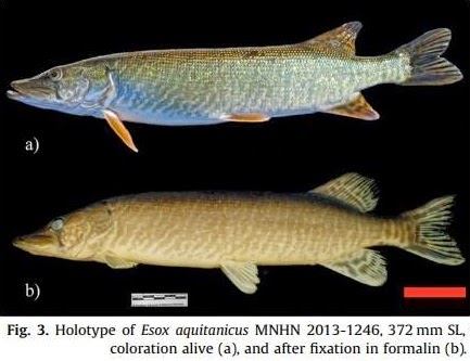
| 5:06p | [Ichthyology • 2014] Esox aquitanicus | Aquitanian pike | brochet aquitain • Morphological and Molecular Evidence of Three Species of Pikes Esox spp. (Actinopterygii, Esocidae) in France, including the Description of A New Species
Abstract This integrative taxonomy study of French pikes compares morphological characters and molecular sequence data (mitochondrial COI and nuclear Plagl2 genes). In addition to the expected E. lucius, DNA sequences and morphology both support a new species in France, E. aquitanicus sp. nov. from the Charente to the Adour drainages. It is characterized by a color pattern of sides with narrow 1–1.5-scale-wide oblique vertical bands, conferring it a marbled coat, a snout only 0.9 times larger than the postorbital length, an anal fin basis 1.1–1.2 times larger than the caudal peduncle length, 101 to 121 lateral scales, 53 to 57 vertebrae, as well as 24 diagnostic sites in the COI gene and 3 in the Plagl2 gene. Partial COI sequences (131 bp) from modern and historical specimens indicate also the presence of E. cisalpinus and E. lucius during the 19th century in Lake Geneva. Morphological and molecular data points to a possible hybridization between E. lucius with both other local pike species, representing a risk for them. Their endangerment status should be evaluated rapidly in order to take conservation measures. Keywords: Esox aquitanicus; Esox cisalpinus; New species; Integrative taxonomy; Cytochrome c oxidase subunit 1; Pleiomorphic adenoma gene-like 2; France Etymology: The specific name aquitanicus is the adjective of Aquitania referring to the region of southwestern France, Aquitaine, where the species was discovered. For this reason, the vernacular name chosen is “Aquitanian pike” (“brochet aquitain” in French). Gaël Pierre Julien Denysa, Agnès Dettaib, Henri Persatc, Mélyne Hautecœura and Philippe Keitha. 2014. Morphological and Molecular Evidence of Three Species of Pikes Esox spp. (Actinopterygii, Esocidae) in France, including the Description of A New Species [Évidence morphologique et moléculaire de trois espèces de brochets Esox spp. (Actinopterygii, Esocidae) en France, dont la description d’une nouvelle espèce] Comptes Rendus Biologies. DOI: dx.doi.org/10.1016/j.crvi.2014.07.002 |
| << Previous Day |
2014/08/16 [Calendar] |
Next Day >> |