[Most Recent Entries] [Calendar View]
Thursday, January 7th, 2016
| Time | Event | ||||||
| 4:29a | [PaleoOrnithology • 2016] Is the “Genyornis” Egg of A Mihirung or Another Extinct Bird from the Australian Dreamtime?
Highlights • Eggshell previously identified as from Genyornis newtoni is reassessed. • Egg size and microstructure is not conducive with an identity as an dromornithid. • We suggest that this eggshell is from one of the extinct megapodes in Progura. • Previous assessments of the timing of Genyornis extinction relate to Progura species. Abstract The iconic Australian Genyornis newtoni (Dromornithidae, Aves) is the sole Pleistocene member of an avian clade now hypothesized to be alternatively in Anseriformes or the sister group of crown Galloanseres. A distinctive type of fossil eggshell commonly found in eroding sand dunes, has been referred to Genyornis newtoni since the 1980s. The 126 by 97 mm Spooner Egg, dated at 54.7 ± 3.1 ka by optical dating of its enclosing sediments, is a complete specimen of this eggshell type that was reconstructed from fragments of a broken egg. We show that the size of the eggs from which this ‘Genyornis’ eggshell derives, either as predicted from measurements of fragments, or as indicated by the Spooner Egg, is unexpectedly small given the size of G. newtoni, which has an estimated mass of 275 kg, or about seven times the mass of the emu that has a similar sized egg. We compared the microstructure of the putative Genyornis eggshell to that of other dromornithids and a range of galloanseriform taxa using several microcharacterisation techniques. The ‘Genyornis’ eggshell displays a mosaic of oological characters that do not unambiguously support referral to any known modern bird. Its shell structure, coupled with chemical compounds in the accessory layer, makes it unlikely to have been laid by a dromornithid, whereas several characters support a megapode origin. A potential candidate for the bird that laid the putative ‘Genyornis’ eggs in the Pleistocene fossil avifaunal record has been ignored: Progura, a genus of extinct giant megapodes, whose species were widespread in Australia. Regression of egg size of megapodes and body mass shows that the Spooner Egg approximates the expected size for eggs laid by species of Progura. We advance the suggestion that the fossil eggshell hitherto referred to Genyornis newtoni, is more likely to have been laid by species of the giant extinct Progura. As megapodes, the species of Progura were obligate ectothermic incubators, which we suggest laid their eggs into a hole dug in sand like the modern megapode Macrocephalon maleo, thus explaining the abundant ‘Genyornis’ eggshell in sand dunes. Referral of this eggshell to Progura means that the fossil record of Genyornis newtoni is limited to bones and the timing of the extinction of this last dromornithid is unknown. In addition, structural similarities of eggshell in megapodes, the putative Genyornis eggshell and dromornithids, raise the possibility that these taxa are phylogenetically more closely related to each other than any is to anseriforms. Specifically, this means that dromornithids might be a sister group to galliforms rather than to or within anseriforms. Keywords: Eggs; Eggshells; Paleoenvironments; Genyornis newtoni; Dromornithids; Megapodes; Progura; Micro-CT; EBSD; Quaternary; Australia Conclusions We have described in detail the structure of putative Genyornis eggshell and raised several obstacles to the hypothesis first advanced by Williams (1981) and accepted thereafter ( Miller et al., 1999 and Miller et al., 2005), that this ootype was laid by the giant dromornithid G. newtoni. Rather, we think it more likely that it was laid by one of the several species of giant megapodes in the genus Progura that were widespread in the Pleistocene in Australia. Gerald Grellet-Tinner, Nigel A. Spooner and Trevor H. Worthy. 2016. Is the “Genyornis” Egg of A Mihirung or Another Extinct Bird from the Australian Dreamtime? QUATERNARY SCIENCE REVIEWS. 133:147-164 DOI: 10.1016/j.quascirev.2015.12.011 | ||||||
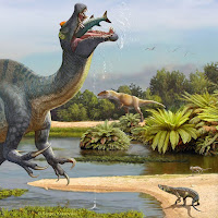
| 4:33p | [Paleontology • 2016] Morphofunctional Analysis of the Quadrate of Spinosauridae (Dinosauria: Theropoda) and the Presence of Spinosaurus and a Second Spinosaurine Taxon in the Cenomanian of North Africa
Abstract Six quadrate bones, of which two almost certainly come from the Kem Kem beds (Cenomanian, Upper Cretaceous) of south-eastern Morocco, are determined to be from juvenile and adult individuals of Spinosaurinae based on phylogenetic, geometric morphometric, and phylogenetic morphometric analyses. Their morphology indicates two morphotypes evidencing the presence of two spinosaurine taxa ascribed to Spinosaurus aegyptiacus and ?Sigilmassasaurus brevicollis in the Cenomanian of North Africa, casting doubt on the accuracy of some recent skeletal reconstructions which may be based on elements from several distinct species. Morphofunctional analysis of the mandibular articulation of the quadrate has shown that the jaw mechanics was peculiar in Spinosauridae. In mature spinosaurids, the posterior parts of the two mandibular rami displaced laterally when the jaw was depressed due to a lateromedially oriented intercondylar sulcus of the quadrate. Such lateral movement of the mandibular ramus was possible due to a movable mandibular symphysis in spinosaurids, allowing the pharynx to be widened. Similar jaw mechanics also occur in some pterosaurs and living pelecanids which are both adapted to capture and swallow large prey items. Spinosauridae, which were engaged, at least partially, in a piscivorous lifestyle, were able to consume large fish and may have occasionally fed on other prey such as pterosaurs and juvenile dinosaurs.  Systematic Paleontology Dinosauria Owen, 1842 Saurischia Seeley, 1887 Theropoda Marsh, 1881 Tetanurae Gauthier, 1986 Megalosauroidea (Fitzinger, 1843) Walker 1964 Spinosauridae Stromer, 1915 Spinosaurinae (Stromer, 1915) Sereno et al., 1998 Description: The six isolated quadrates from the Kem Kem beds of Morocco clearly belong to two morphotypes (Figs 2–4) based on the size and outline of the quadrate foramen, shape of the mandibular articulation, and outline, surface, and orientation of the quadratojugal contacts. Measurements taken on each quadrate (Fig 5A–5D) are provided in Table 1. Spinosaurus Stromer, 1915 Spinosaurus aegyptiacus Stromer, 1915  ?Sigilmassasaurus Russel, 1996 ?Sigilmassasaurus brevicollis Russel, 1996
Conclusion The description and identification of six isolated quadrates, among which two most probably come from the Kem Kem beds of Morocco, provide additional information on the Cenomanian dinosaur fauna of North Africa. Based on cladistic, geometric morphometric, and phylogenetic morphometric analyses, two morphotypes have been successfully identified as belonging to two species of Spinosaurinae, and ascribed to Spinosaurus aegyptiacus and ?Sigilmassasaurus brevicollis. This study provides the first convincing evidence of two spinosaurine taxa in the Cenomanian of North Africa based on cranial material, casting doubt on the recent reconstruction of a quadrupedal Spinosaurus which may be based on individuals belonging to two different species of Spinosaurinae. Ontogenetic changes occurring in the spinosaurid quadrates include the suture of the quadrate and quadratojugal, delimitation of the mandibular condyles and squamosal capitulum, and development of a ventral projection of the dorsal quadratojugal contact and a second quadrate ridge ventral to the quadrate head. Based on the quadrate proportions and estimated skull length of Baryonyx and Spinosaurus, quadrates of mature individuals from Morocco belong to animals with a skull length of no more than 120 cm. This suggests that very large forms of Spinosaurus may have been rare in the Kem Kem assemblages. Morphofunctional analysis of the spinosaurid quadrates has revealed peculiar jaw mechanics in these specialized theropods. An helicoidal and strongly lateromedially oriented joint of the jaw articulation allowed the lateral displacement of the mandibular ramus when the lower jaw was depressed. This lateral movement of the ramus was possible due to a movable mandibular symphysis as the dentaries were joined by connective tissues, and allowed the pharynx to be widened. A similar jaw articulation was convergently present in pterosaurs and particularly pelecanids which also have a mandibular symphysis restricted to the anterior extremity of the mandible. Spinosauridae, which are considered to be semi-aquatic and partially piscivorous animals, were able to swallow large prey such as fish in the same way as pelecanids.  
Christophe Hendrickx , Octávio Mateus and Eric Buffetaut. 2016. Morphofunctional Analysis of the Quadrate of Spinosauridae (Dinosauria: Theropoda) and the Presence of Spinosaurus and a Second Spinosaurine Taxon in the Cenomanian of North Africa. PLoS ONE. 11(1): e0144695. DOI: 10.1371/journal.pone.0144695 |


| << Previous Day |
2016/01/07 [Calendar] |
Next Day >> |