[Most Recent Entries] [Calendar View]
Sunday, March 1st, 2020
| Time | Event | ||||||
| 8:54a | [Herpetology • 2020] Kurixalus gracilloides • A New Species of Kurixalus (Anura: Rhacophoridae) from northern Vietnam with Comments on the Biogeography of the Genus
ABSTRACT We describe a new species of rhacophorid frogs from Nghe An Province in northern Vietnam based on morphological and molecular evidences. Morphologically, Kurixalus gracilloides sp. nov. is distinguished from its congeners by a combination of the following diagnostic characters: body size small (snout–vent length 27.9–31.2 mm in males); head width subequal to head length; snout rounded with no dermal projection; canthus rostralis distinct, curved; vomerine teeth present; single internal vocal sac; iris golden-brown; small nuptial pad in finger I; dorsal surfaces golden-brown with a saddle-shaped dark marking; large dark spots on ventral surfaces absent; dermal fringes along outer edge of limbs; conical dermal appendage at the heel; skin on dorsum rough; skin on throat and chest granular; finger webbing rudimentary and toe webbing moderately developed, webbing formula I 2–2½ II 1½–3 III 1¾–3½ IV 3–1½ V. The new species is separated from all other congeners by uncorrected genetic distances ranging from 5.4% to 12.7% based on mitochondrial 16S rRNA gene. Phylogenetic analyses of mtDNA suggest that the new species is nested within a clade of Taiwanese and Yunnan Kurixalus with strong support values. The new species is currently known only from secondary bamboo forest in Pu Mat National Park, northern Vietnam, at elevations of 150 m asl. We suggest the new species should be considered as Near Threatened (NT) following the IUCN’s Red List categories. KEYWORDS: Kurixalus gracilloides sp. nov., Gracixalus, Pu Mat National Park, Nghe An Province, morphology, mtDNA, 16S rRNA
Family RHACOPHORIDAE Hoffman, 1932 Genus Kurixalus Ye, Fei, & Dubois in Fei, 1999 Kurixalus gracilloides sp. nov. Etymology: The specific epithet ‘gracilloides’ is a Latin adjective in nominative singular, derived from Latin ‘gracilis’ (gracile, thin) and ‘-oides’ (similar to, resembling something). The species name is given to a remarkable similarity of the new species with several species of the genus Gracixalus in body shape and dorsum colouration. Recommended vernacular names: We suggest the following common names: Gracile Frilled Treefrog (English), Stroynyi Bakhromchatyi Veslonog (Russian) and Ếch cây rìa chân mảnh (Vietnamese). Tan Van Nguyen, Tang Van Duong, Kien Trung Luu and Nikolay A. Poyarkov. 2020. A New Species of Kurixalus (Anura: Rhacophoridae) from northern Vietnam with Comments on the Biogeography of the Genus. Journal of Natural History. DOI: 10.1080/00222933.2020.1728411  | ||||||
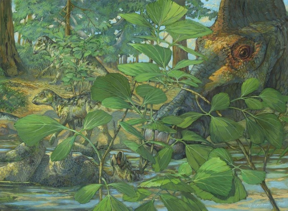
| 2:29p | [Paleontology • 2020] Evidence of Proteins, Chromosomes and Chemical Markers of DNA in Exceptionally Preserved Dinosaur Cartilage
Abstract A histological ground-section from a duck-billed dinosaur nestling (Hypacrosaurus stebingeri) revealed microstructures morphologically consistent with nuclei and chromosomes in cells within calcified cartilage. We hypothesized that this exceptional cellular preservation extended to the molecular level and had molecular features in common with extant avian cartilage. Histochemical and immunological evidence supports in situ preservation of extracellular matrix components found in extant cartilage, including glycosaminoglycans and collagen type II. Furthermore, isolated Hypacrosaurus chondrocytes react positively with two DNA intercalating stains. Specific DNA staining is only observed inside the isolated cells, suggesting endogenous nuclear material survived fossilization. Our data support the hypothesis that calcified cartilage is preserved at the molecular level in this Mesozoic material, and suggest that remnants of once-living chondrocytes, including their DNA, may preserve for millions of years. Keywords: cartilage, dinosaur, nuclei, chromosomes, collagen II, DNA markers
  CONCLUSIONS: The identification of chemical markers of DNA in Hypacrosaurus suggest it may preserve much longer than originally proposed [30,31]. Even though it is clear that contamination does exist in fossil material and complicates identifications of original organic molecules, it can be accounted for with proper controls. Contamination is not a plausible explanation in this case, and to this date, the possible preservation of original proteins and DNA in deep time has not been convincingly eliminated with data. Although extensive research and sequencing is required to further understand DNA preservation in Mesozoic material, along with its chemical and molecular alterations, our data suggest the preserved nuclear material in Hypacrosaurus was in a condensed state at the time of the death of the organism, which may have contributed to its stability. We propose that DNA condensation may be a favorable process to its fossilization. Additionally, as was suggested for protein fossilization [20,45,46], crosslinking may be another mechanism involved in the preservation of DNA in deep time. Alida M. Bailleul, Wenxia Zheng, John R. Horner, Brian K. Hall, Casey M. Holliday and Mary H. Schweitzer. 2020. Evidence of Proteins, Chromosomes and Chemical Markers of DNA in Exceptionally Preserved Dinosaur Cartilage. National Science Review. nwz206. DOI: 10.1093/nsr/nwz206   | ||||||
| 3:05p | [Botany • 2020] Primulina inflata (Gesneriaceae) • A New Species of Primulina from Danxia landform in Jiangxi, China
Abstract Based on morphological observations and comparisons, a new species of Primulina (Gesneriaceae), Primulina inflata Li.H. Yang & M.Z. Xu, is described and illustrated. This new species resembles P. xiuningensis in leaf blade shape and indumentum, and differs from the latter by the white corolla with longitudinally purple-red stripes (vs. yellowish without stripes), inflated tubular corolla tube (vs. tubular) and straight filament (vs. geniculate). The descriptions, illustrations and photographs of this new species are provided here. Keyword: China, Gesneriaceae, Primulina, Primulina inflata, P. xiuningensis, Taxonomy

Primulina inflata Li.H. Yang & M.Z. Xu, sp. nov. 粗筒小花苣苔 Diagnosis: Primulina inflata differs from P. xiuningensis by the white corolla with longitudinally purple-red stripes (vs. yellowish without stripes), inflated corolla tube (vs. tubular) and straight filaments (vs. geniculate). Distribution and habitat: As most other Primulina species, P. inflata is a species endemic to Xingguo County, Jiangxi Province. Based on our field investigations, this new species is only found at its type locality at Bingxindong scenic area in a Danxia Geopark. Plants of this new species grow on the moist rock surface of a cave entrance. We observed about 200 mature individuals of this new species at the type locality in 2016, but no more than 50 mature individuals in 2018. The serious reduction of the population size mostly resulted from human disturbance. Based on this information, P. inflata is considered as Critically Endangered (CR): B2a,b(iii,v); C2a(i), following the IUCN categories and criteria (IUCN 2016). However, our field investigations on this species are insufficient, and more fieldwork is required to comprehensively understand its geographical distribution. Etymology: The specific epithet is derived from the inflated corolla tube of this new species. Mei-Zhen Xu, Hang-Hui Kong, Ming Kang and Li-Hua Yang. 2020. A New Species of Primulina (Gesneriaceae) from Danxia landform in Jiangxi, China. Taiwania. 65(2); 163-166.     |


| << Previous Day |
2020/03/01 [Calendar] |
Next Day >> |