[Most Recent Entries] [Calendar View]
Sunday, July 25th, 2021
| Time | Event | ||||||
| 3:36a | [Herpetology • 2021] First National Record of Gracixalus quangi and G. yunnanensis (Anura: Rhacophoridae) from Thailand
Abstract Background: The bushfrog genus Gracixalus Delorme, Dubois, Grosjean & Ohler, 2005 is found in southern and south-western China, Vietnam, Laos, Thailand and Myanmar. It is presently comprised of 17 species. In Thailand, only two species have been recorded, namely G. carinensis (Boulenger) and G. seesom (Massui, Khonsue, Panha & Eto). The latter of these two species is currently known to be endemic to the country. New information: Based on recent field work conducted in 2019 in Doi Phu Kha National Park, Nan Province of northern Thailand, we are reporting two new records of the genus Gracixalus, G. quangi and G. yunnanensis, from Thailand, based on morphological and molecular evidence. In addition, this is the first study to report on the identification of a female specimen of G. yunnanensis. Furthermore, morphological data and natural history notes of the aforementioned species in Thailand have been provided, along with updated locations for the distribution of both species. Keywords: Gracixalus quangi, G. yunnanensis, new record, 16s rRNA, Nan Province
Sengvilay Lorphengsy, Tan Van Nguyen, Nikolay A. Poyarkov, Yun-He Wu, Parinya Pawangkhanant, Supaporn Passorn, Jing Che and Chatmongkon Suwannapoom. 2021. First national record of Gracixalus quangi Rowley, Dau, Nguyen, Cao & Nguyen, 2011 and G. yunnanensis Yu, Li, Wang, Rao, Wu & Yang, 2019 (Amphibia: Anura: Rhacophoridae) from Thailand. Biodiversity Data Journal. 9: e67667. DOI: 10.3897/BDJ.9.e67667 | ||||||
| 2:39p | [Botany • 2021] Amomum fangdingii (Zingiberaceae) • A New Species from Guangxi, China [Taxonomic Studies on Amomum in China III]
Abstract A plant formerly misidentified as Amomum maximum in Guangxi, China, is described here as a new species, namely A. fangdingii. A detailed description, distribution map and illustrations are provided together with a taxonomic key distinguishing A. fangdingii from eight morphologically similar species characterised by cincinnate inflorescences occurring in Cambodia, China, Laos and Vietnam. Keywords: Amomum maximum, A. velutinum, gingers, misidentification, Monocots.
Amomum fangdingii X.E.Ye, Škorničk. & N.H.Xia sp. nov. Xing-Er Ye, Jana Leong-Škorničková, Lin Bai and Nian-He Xia. 2021. Taxonomic Studies on Amomum (Zingiberaceae) in China III: Amomum fangdingii, A New Species from Guangxi. Phytotaxa. 490.3; 263–270. DOI: 10.11646/phytotaxa.490.3.4 | ||||||
| 2:41p | [Mollusca • 2021] Snails Riding Mantis Shrimps: Ectoparasites evolved from Ancestors Living as Commensals on the Host’s Burrow Wall
Highlights • Snails of the genus Caledoniella are highly adapted ectoparasites on mantis shrimps. • They originated from commensal ancestors that lived on the host burrow wall. • Contrary to previous classifications, Caledoniella is placed within Truncatelloidea. • Five families, including Caledoniellidae, are redefined in Truncatelloidea. • Symbiotic mode of life has evolved multiple times in this superfamily. Abstract The molluscan class Gastropoda includes over 5,000 parasitic species whose evolutionary origins remain poorly understood. Marine snails of the genus Caledoniella (Caledoniellidae) are obligate parasites that live on the abdominal surface of the gonodactylid mantis shrimps. They have highly modified morphological characteristics specialized to the ectoparasitic lifestyle that make it difficult to infer their close relatives, thereby posing a question about their current systematic position in the superfamily Vanikoroidea. In the present study, we performed molecular phylogenetic analyses using three nuclear and three mitochondrial gene sequences to unveil the phylogenetic position of these enigmatic snails. The resulting trees recovered Caledoniella in the superfamily Truncatelloidea and within a subclade of commensal species that live on the burrow wall of marine benthic invertebrates. More specifically, Caledoniella formed the sister clade to a commensal snail species living in mantis-shrimp burrows and they collectively were sister to Sigaretornus planus (formerly in the family Tornidae or Vitrinellidae), a commensal living in echiuran burrows. This topology suggests that the species of Caledoniella achieved their ectoparasitic mode of life through the following evolutionary pathway: (1) invasion into the burrows of benthic invertebrates, (2) specialization to mantis shrimps, and (3) colonization of the host body surface from the host burrow wall with the evolution of the parasitic nature. The final step is likely to have been accompanied by the acquisition of a sucker on the metapodium, the loss of the radula and operculum, and the formation of monogamous pair bonds. The present molecular phylogeny also suggested parallel evolution of planispiral shells in a subclade of Truncatelloidea and enabled us to newly redefine the families Caledoniellidae, Elachisinidae, Teinostomatidae, Tornidae and Vitrinellidae. Keywords: Adaptation, Caledoniella, Parasitism, Symbiosis, Stomatopoda, Truncatelloidea
 Ryutaro Goto, Tsuyoshi Takano, Douglas J. Eernisse, Makoto Kato and Yasunori Kano. 2021. Snails Riding Mantis Shrimps: Ectoparasites evolved from Ancestors Living as Commensals on the Host’s Burrow Wall. Molecular Phylogenetics and Evolution. 163, 107122. DOI: 10.1016/j.ympev.2021.107122 | ||||||
| 2:42p | [Paleontology • 2021] Osteological Revision of the Holotype of the Middle Jurassic Sauropod Dinosaur Patagosaurus fariasi Bonaparte, 1979 (Sauropoda: Cetiosauridae)
Middle Jurassic sauropod taxa are poorly known, due to a stratigraphic bias of localities yielding body fossils. One such locality is Cerro Cóndor North, Cañadón Asfalto Formation, Patagonia, Argentina, dated to latest Early−Middle Jurassic. From this locality, the holotype of Patagosaurus fariasi Bonaparte 1986 is revised. The material consists of the axial skeleton, the pelvic girdle, and the right femur. Patagosaurus is mainly characterised by a combination of features mainly identified on the axial skeleton, including the following: 1) cervical centra with low Elongation Index; 2) high projection of the postzygodiapophyseal lamina; 3) deep anterior pleurocoels that are sometimes compartmentalized in cervicals; 4) high projection of the neural arch and spine in dorsal vertebrae and anterior(most) caudal vertebrae; 5) deep pneumatic foramina in posterior dorsals which connect into an internal pneumatic chamber; and 6) anterior caudal vertebrae with ‘saddle’ shaped neural spines. Diagnostic features on the appendicular skeleton include: 1) a transversely wide and anteroposteriorly short femur; 2) a medial placement of the fourth trochanter on the femur; and 3) an anteroposteriorly elongated ilium with a rounded dorsal rim, with hook-shaped anterior lobe. The characters that are diagnostic for Patagosaurus are discussed, and the osteology of Patagosaurus is compared to that of Early and Middle Jurassic (eu)sauropods from both Laurasia and Gondwana. KEYWORDS: Sauropoda, Eusauropoda, Patagosaurus, Gondwana, Middle Jurassic, Patagonia, pneumaticity 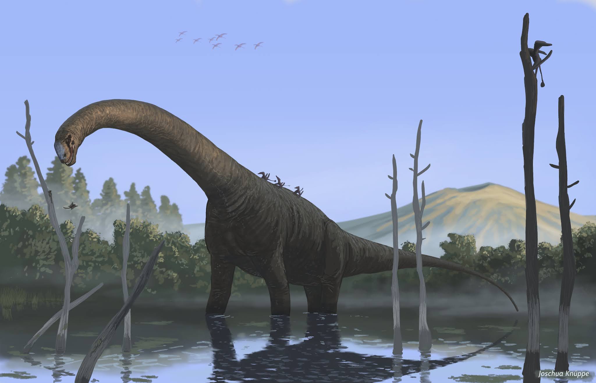 Femke M. Holwerda, Oliver W. M. Rauhut and Diego Pol. 2021. Osteological Revision of the Holotype of the Middle Jurassic Sauropod Dinosaur Patagosaurus fariasi Bonaparte, 1979 (Sauropoda: Cetiosauridae). Geodiversitas. 43 (16); 575-643. DOI: 10.5252/geodiversitas2021v43a16. geodiversitas.com/43/16 |


| << Previous Day |
2021/07/25 [Calendar] |
Next Day >> |