[Recent Entries][Archive][Friends][User Info]
September 10th, 2011
| September 10th, 2011 | |
|---|---|
| 06:36 pm [industrialterro] [Link] |
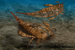
Acutiramus Acutiramus is an extinct genus of eurypterid which lived in the Late Silurian (Ludlow) to Early Devonian. Acutiramus was one of the largest eurypterids with pincers 5 cm and length about 2 m. It was related to another large eurypterid, Pterygotus. Pterygotidae, which lived from the Ordovician to Devonian periods, were characterized by small to large exoskeletons with semilunar scales. The telson, (tail) was expanded, or flatter than it was tall. Pterygotidae also had chelicerae (claws in front of the mouth) that were large and long, with strong, well developed teeth on the claws. Their walking legs were small and slender, without spines. Acutiramus is distinguishable from other Pterygotidae by the distal margin of the chelae, where the final tooth is at an acute angle to the rest of the claw (hence the name Acutiramus, or “acute arm”). The large tooth in the center of the claw is distally inclined, which is to say it points forwards. The prosoma (head) is subquadrate, with compound eyes located at the edge of the front corners. The telson has a low row of knobs running down its center. Ричард Лауб (Музей науки Буффало), Виктор Толлертон (Музей штата Нью-Йорк) и Ричард Беркоф (Технологический институт Стивенса) сумели показать, что механические ограничения клешни ракоскорпиона Acutiramus не позволяли ему пробивать внешнюю оболочку мечехвоста средних размеров без вреда для себя. Отмечается также, что отсутствие аналога локтевого сустава между клешней и телом ракоскорпиона ограничивало его движения. Это позволяло животному более эффективно ловить добычу, лежащую на морском дне, чем цепляться за быстро плывущих рыб и прочих существ. Зубчатые шипы, которыми были усеяны клешни, вероятно, помогали ему хватать и кромсать жертву, но как активный хищник Acutiramus действовать не мог. Скорее всего, он был падальщиком или даже вегетарианцем.
Ископаемые останки (1, 2, 3, 4):
Tags: Вымершие членистоногие, Силур |
| Time | Event |
| 06:53 pm [industrialterro] [Link] |
Slimonia Slimonia is a genus of Silurian eurypterid roughly similar to the genus Pterygotus. Slimonia resembled Pterygotus, save that the former's telson is larger, and that its body was smaller and more slender than the latter. Unlike Pterygotus, which lived in estuaries, Slimonia species lived exclusively in freshwater environments. Slimonia preyed on smaller fish, such as heterostracans and early osteostracans by seizing and rending them with its large chelicerae. The largest species of Slimonia was extremely long, around two meters. It carried its body on spindly legs and was likely an ambush predator. The lungs of the species were located on the underside of the body in a series of folds. Slimonia is distinguishable by its quadrate (roughly square) prosoma, or head, with small compound eyes on the front corners. They had large cordate bodies, with a narrow postabdomen and a telson with a "strongly expanded anterior half." Their chelicerae (claws) were small; their walking legs had denticles, but no spines. Genital appendages were long and narrow in both males and females.
Tags: Вымершие членистоногие, Силур |
| Time | Event |
| 07:16 pm [industrialterro] [Link] |
Mixopterus Mixopterus is a genus of a eurypterid. It belongs to the family Mixopteridae. Mixopterus were characterised by a robust exoskeletons with scattered tubercles or semicircular scales. The prosoma (head) was subquadrate, protruding antemedially. The chelicerae (claws in front of the mouth) were small. The first two pairs of walking legs were strongly developed, with long paired spines. The third and fourth walking legs were moderately sized, with short spines. The preabdomen, the front portion of the body, was narrow with axial furrows, while the postabdomen was narrow. The telson was a curved spine.
Ископаемые останки (1, 2, 3, 4):
Tags: Вымершие членистоногие, Силур |
| Time | Event |
| 07:43 pm [industrialterro] [Link] |
Eurypterus Эвриптерус (Eurypterus) имел шесть пар конечностей: первая пара – хелицеры, следующие четыре пары – короткие шипастые, используемые для хождения, и шестая пара выступает наружу в виде лопат и весел. Хелицеры были столь малы, что ходильные конечности должны были использоваться для удержания пищи, а основания лопатовидных конечностей, несущие зубцы, служили для перетирания. Последняя пара (шестая) конечностей просомы были самые крупные и на концах сильно расширенными; они, по-видимому, служили для плавания. Два крупных сложных глаза и два маленьких простых находились на спинной стороне просомы. E. remipes are usually between 5 to 8 in (13 to 20 cm) in length. E. lacustris average at larger sizes at 6 to 9 in (15 to 23 cm) in length. The largest specimen of E. remipes ever found was 1.3 m (4.3 ft) long, currently on display at the Paleontological Research Institution of New York. Eurypterus fossils often occur in similar sizes in a given area. This may be a result of the fossils being 'sorted' into windrows as they were being deposited in shallow waters by storms and wave action. The Eurypterus body is broadly divided into two parts: the prosoma and the opisthosoma (in turn divided into the mesosoma and the metasoma). The prosoma is the forward part of the body, it is actually composed of six segments fused together to form the head and the thorax. It contains the semicircular to subrectangular platelike carapace. On the dorsal side of the latter are two large crescent-shaped compound eyes. They also possessed two smaller light-sensitive simple eyes (the median ocelli) near the center of the carapace on a small elevation (known as the ocellar mound). Underneath the carapace is the mouth and six appendages, usually referred to in Roman numerals I-VI. Each appendage in turn is composed of nine segments (known as podomeres) labeled in Arabic numerals 1-9. The first segments which connect the appendages to the body are known as the coxa (plural coxae). The first pair (Appendage I) are the chelicerae, small pincer-like arms used for tearing food apart (mastication) during feeding. After the chelicerae are three pairs of short legs (Appendages II, III, and IV). They are spiniferous, with predominantly two spines on each podomere and with the tipmost segment having a single spine. The last two segments are often indistinguishable and give the appearance of a single segment having three spines. They are used both for walking and for food capture. The next pair (Appendage V) is the most leg-like of all appendages, longer than the first three pairs and are mostly spineless except at the tipmost segments. The last pair (Appendage VI) are two broad paddle-like legs used for swimming. The coxae of Appendage VI are broad and flat, resembling an 'ear'. The ophisthosoma (the abdomen) is composed of 12 segments, composed of fused upper plates (tergites) and bottom plates (sternites). It is further subdivided in two ways. Based on the width and structure of each segment, they can be divided into the broad preabdomen (segments 1 to 7) and the narrow postabdomen (segments 8 to 12). The preabdomen is the broader segments of the anterior portion of the ophisthosoma while the postabdomen are the last five segments of the Eurypterus body. Each of the segments of the postabdomen contain lateral flattened protrusions known as the epimera with the exception of the last needle-like (styliform) segment known as the telson (the 'tail'). The segment immediately preceding the telson (which also has the largest epimera of the postabdomen) is known as the pretelson. An alternative way to divide the ophisthosoma is by function. It can also be divided into the mesosoma (segments 1 to 6), and the metasoma (segments 7 to 12). The mesosoma contains the gills and reproductive organs of Eurypterus. Its ventral segments are overlaid by appendage-derived plates known as Blatfüsse (singular Blatfuss, German for "sheet foot"). Protected within which are the branchial chambers which contain the respiratory organs of Eurypterus. The metasoma, meanwhile, do not possess Blatfüsse. Some authors incorrectly use mesosoma and preabdomen interchangeably, as with metasoma and postabdomen. The main respiratory organs of Eurypterus were what seems to be book gills, located in branchial chambers within the segments of the mesosoma. They may have been used for underwater respiration. They are composed of several layers of stacked thin tissue resembling the pages of a book, hence the name. In addition, they also possessed five pairs of oval-shaped areas covered with microscopic projections on the ceiling of the second branchial chambers within the mesosoma, immediately below the gill tracts. These areas are known as Kiemenplatten (or gill-tracts, though the former term is preferred). They are unique to eurypterids. Eurypterus are sexually dimorphic. On the bottom side of the first two segments of the mesosoma are central appendages used for reproduction. In females, they are long and narrow. In the males they are very short.[23] A minority of authors, however, assume the reverse: longer genital appendage for males, shorter for females. The exoskeleton of Eurypterus is often covered with small outgrowths known as ornamentation. They include pustules (small protrusions), scales, and striations.They vary by species and are used for identification. For more detailed diagnostic descriptions of each species under Eurypterus, see sections below.
Способ перемещения:
Размеры в сравнении с человеком:
Ископаемые останки (1, 2, 3, 4):
Tags: Вымершие членистоногие, Силур |


_buffaloenisis.bmp)


| Previous Day | 2011/09/10 [Archive] |
Next Day |